Home
/ Cross Section Of A Bone Labeled - The Parts Of A Healthy Long Bone With A Cross Section Showing The Inside Of The Bone Basic Anatomy And Physiology Anatomy And Physiology Anatomy : This category is largely categorized by the content of the bone rather than the shape.
Cross Section Of A Bone Labeled - The Parts Of A Healthy Long Bone With A Cross Section Showing The Inside Of The Bone Basic Anatomy And Physiology Anatomy And Physiology Anatomy : This category is largely categorized by the content of the bone rather than the shape.
Cross Section Of A Bone Labeled - The Parts Of A Healthy Long Bone With A Cross Section Showing The Inside Of The Bone Basic Anatomy And Physiology Anatomy And Physiology Anatomy : This category is largely categorized by the content of the bone rather than the shape.. The surface features of bones vary considerably, depending on the function and location in the body. The diaphysis and the epiphysis. The strength, shape and stability of the human body are dependent on the musculoskeletal system. Sketch and label of a cross section of a long bone : There are 3 types of bone tissue, including the following:
The outlined area is a cross section of an osteon of compact bone. Forms the larger rounded ends of long bones. This photo shows a cross section through bone. Area between the diaphysis and epiphysis at both ends of the bone. Bone test anatomy and physiology 12 photos of the bone test anatomy and physiology anatomy and physiology bone lab test, anatomy and physiology bone markings test, anatomy and physiology bone practical test, anatomy and physiology bone tissue test, anatomy and physiology test on bone tissue, bone, anatomy and.
I Pinimg Com 564x 03 04 83 0304831f8780c111baaf from i.pinimg.com Trust pharmacy 221 massachusetts ave, boston, ma 02115. It can be found under the periosteum and in the diaphyses of long bones, where it provides support and protection. This diagram depicts cross section human body hip thigh.human anatomy diagrams show internal organs, cells, systems, conditions, symptoms and sickness information and/or tips for healthy living. The strength, shape and stability of the human body are dependent on the musculoskeletal system. Compact bone is the denser, stronger of the two types of bone tissue ( (figure) ). The periosteum contains many strong collagen fibers that are used to firmly anchor tendons and muscles to the bone for movement. The most robust aspect of this unit is the underlying bony architecture. The harder, outer tissue of bones.
It includes such bones as the hip and vertebrae.
Concentric layers of bone cells (osteocytes) and bone matrix surround the central canal. At this level of magnification, the fundamental structure of compact bone is visible. It can be found under the periosteum and in the diaphyses of long bones, where it provides support and protection. This slide contained a cross section of a very small bone, and you are looking at the entire thickness of the shaft of the bone. Related posts of cross section of human bone diagram bone in arm pictures. Related posts of bone cross section labeled bone test anatomy and physiology. This diagram depicts cross section human body hip thigh.human anatomy diagrams show internal organs, cells, systems, conditions, symptoms and sickness information and/or tips for healthy living. Trust pharmacy 221 massachusetts ave, boston, ma 02115. It includes such bones as the hip and vertebrae. Area between the diaphysis and epiphysis at both ends of the bone. They are obtained by taking imaginary slices perpendicular to the main axis of organs, vessels, nerves, bones, soft tissue, or even the entire human body. Forms the larger rounded ends of long bones. Chapter 6 bones and skeletal tissues flashcards quizlet.
They are obtained by taking imaginary slices perpendicular to the main axis of organs, vessels, nerves, bones, soft tissue, or even the entire human body. Cross section of a bone : Cookies allow us to analyze and store information such as the characteristics of your device as well as certain personal data (e.g., ip addresses, navigation, usage or geolocation data, unique identifiers). Imaios and selected third parties, use cookies or similar technologies, in particular for audience measurement. The harder, outer tissue of bones.
Structure Of Bones Longitudinal Section Diagram Quizlet from o.quizlet.com Related posts of bone cross section labeled bone test anatomy and physiology. They are obtained by taking imaginary slices perpendicular to the main axis of organs, vessels, nerves, bones, soft tissue, or even the entire human body. Compact bone is the denser, stronger of the two types of bone tissue ( (figure) ). It includes such bones as the hip and vertebrae. Related posts of cross section of human bone diagram bone in arm pictures. Cross section of a bone : This article lists a series of labeled imaging anatomy cases by system and modality. Concentric layers of bone cells (osteocytes) and bone matrix surround the central canal.
This photo shows a cross section through bone.
Forms the larger rounded ends of long bones. The central haversian canal, and horizontal canals (perforating/ volkmann's) canals contain blood vessels and nerves from the periosteum. This slide contained a cross section of a very small bone, and you are looking at the entire thickness of the shaft of the bone. Plates of cartilage, also known as growth plates which allow the long bones to grow during childhood. They are obtained by taking imaginary slices perpendicular to the main axis of organs, vessels, nerves, bones, soft tissue, or even the entire human body. Sketch and label of a cross section of a long bone : Bones are made of active, living cells that are busy growing, repairing themselves, and communicating Browse 75 cortical bone stock photos and images available, or search for trabecular bone or bone marrow to find more great stock photos and pictures. A long bone has two parts: 100x first focus in the compact decalcified bone (cb) on the left part of the image, you can see small dots, which are. Related posts of cross section of human bone diagram bone in arm pictures. Bone test anatomy and physiology 12 photos of the bone test anatomy and physiology anatomy and physiology bone lab test, anatomy and physiology bone markings test, anatomy and physiology bone practical test, anatomy and physiology bone tissue test, anatomy and physiology test on bone tissue, bone, anatomy and. Bone matrix and cells bone matrix osseous tissue is a connective tissue and like all connective tissues contains relatively few cells and large amounts of extracellular matrix.
To the left is muscle tissue, and to the right is bone marrow. The outlined area is a cross section of an osteon of compact bone. Related posts of bone cross section labeled bone test anatomy and physiology. Forms the larger rounded ends of long bones. On the proximal end of the femur, there are two growth plates.
Bone Structure Anatomy Explained What Is Bone Marrow from www.teachpe.com The previous image was correct, with one between the diaphysis and the head of the femur (which is an ossification center) and the other between the greater trochanter and the diaphysis. The diaphysis is the tubular shaft that runs between the proximal and distal ends of the bone. During the intervening years, there has been little to add to our views as to the best management of acetabular. Bone basics and bone anatomyhave you ever seen fossil remains of dinosaur and ancient human bones in textbooks, television, or in person at a museum? A long bone has two parts: Eliminate sudden changes of direction and influx of one stream into another. Cross section of a bone : Bone in arm pictures 12 photos of the bone in arm pictures bone cancer arm pictures, pictures of bone cancer in arm, bone, bone cancer arm pictures, pictures of bone cancer in arm
The structure of a long bone allows for the best visualization of all of the parts of a bone ( figure 6.7 ).
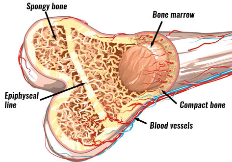
The outside of a bone is covered in a thin layer of dense irregular connective tissue called the periosteum. Bone basics and bone anatomyhave you ever seen fossil remains of dinosaur and ancient human bones in textbooks, television, or in person at a museum? It can be found under the periosteum and in the diaphyses of long bones, where it provides support and protection. The surface features of bones vary considerably, depending on the function and location in the body. It includes such bones as the hip and vertebrae. The most robust aspect of this unit is the underlying bony architecture. Dr calum worsley and assoc prof craig hacking et al. Cross section of a human bone showing bone marrow, spongy bone and blood vessels. Label the haversian canal, osteocyte (mature bone cell) in lacuna, and canaliculi. And recall anatomic structures in cross section. The periosteum contains many strong collagen fibers that are used to firmly anchor tendons and muscles to the bone for movement. This photo shows a cross section through bone. The only section of the proximal end of the femur that articulates is the head.
Each of these cylinders is called an osteon or cross section of a bone. Chapter 6 bones and skeletal tissues flashcards quizlet.

